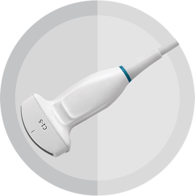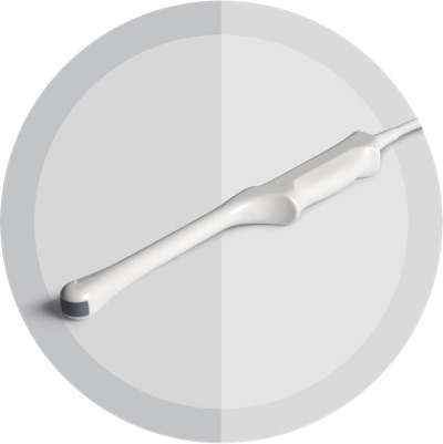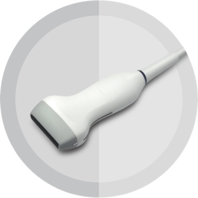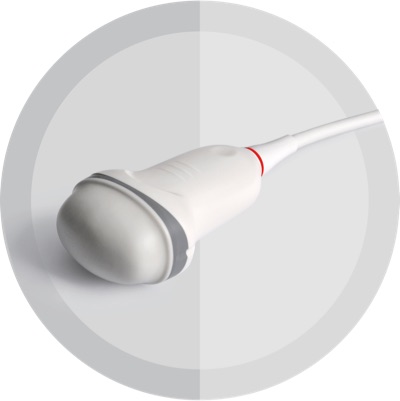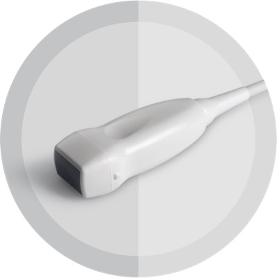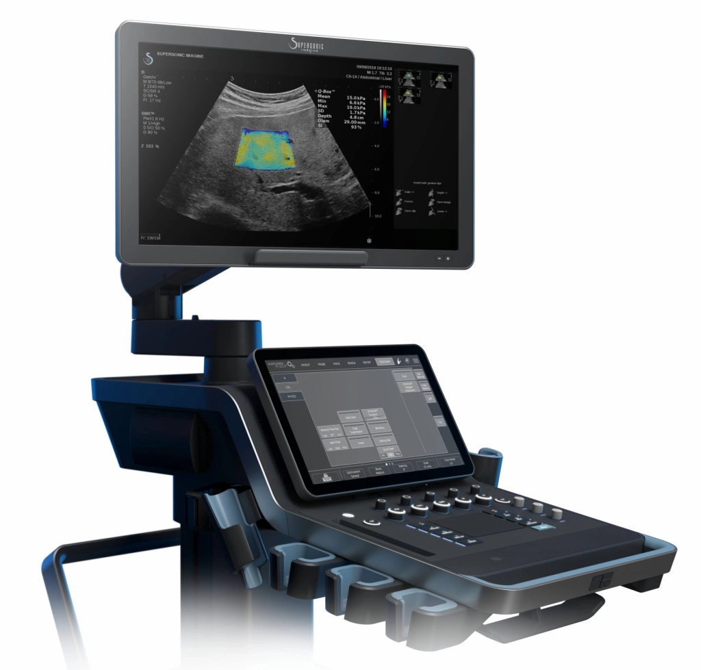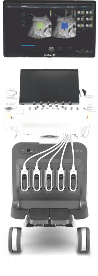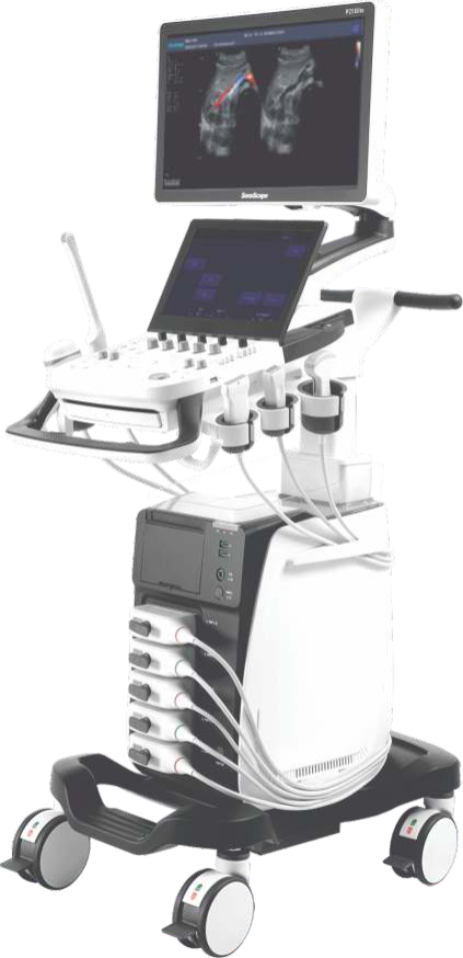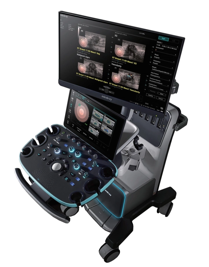



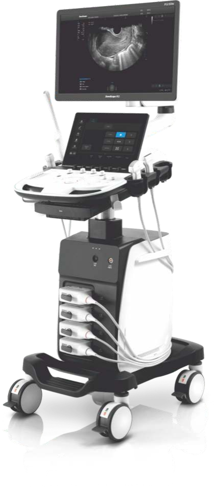
With comprehensive features and optimized probe configurations, P12 Elite provides an exceptional capability to manage all-aspects of the daily clinical practices that help build trust between doctors and patients.
Extraordinary Image Technologies
With the outstanding clear view provided by SonoScape's advanced imaging technologies and optimized transducers, you can make clinical decisions with greater confidence.
μ-Scan+
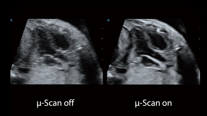
The new generation μ-Scan+ imaging technology greatly improves the visibility of organs and lesions. The high-definition contrast resolution will suppress speckle artifacts while maintaining real tissue architecture.
SR Flow
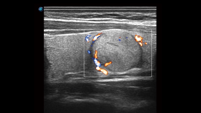
As a new innovative technology, SR Flow improves the capability of detecting low velocity flow signals. It also improves on spatial resolution and overcomes overflow to present users with real hemodynamic information.
μ-Scan+
The new generation μ-Scan+ imaging technology greatly improves the visibility of organs and lesions. The high-definition contrast resolution will suppress speckle artifacts while maintaining real tissue architecture.
SR Flow
As a new innovative technology, SR Flow improves the capability of detecting low velocity flow signals. It also improves on spatial resolution and overcomes overflow to present users with real hemodynamic information.
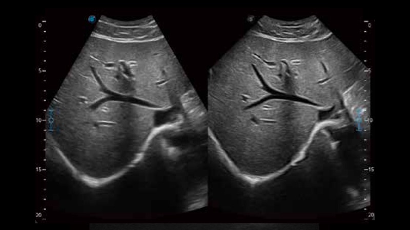
WideScan
With WideScan, the ultrasound image can be enlarged when performing a real time scan by using linear or convex probes, for a more complete view of large lesions or anatomic structures.

Real-time Color Panoramic
With the combination of color flow and real-time panoramic, visualizing the blood flow of an entire vein or artery is now an easy task. Accomplished in real-time for the convenience of the sonographers, any mistake can also be easily back tracked and corrected without interrupting the scan.
Design with Clinical Application in Mind
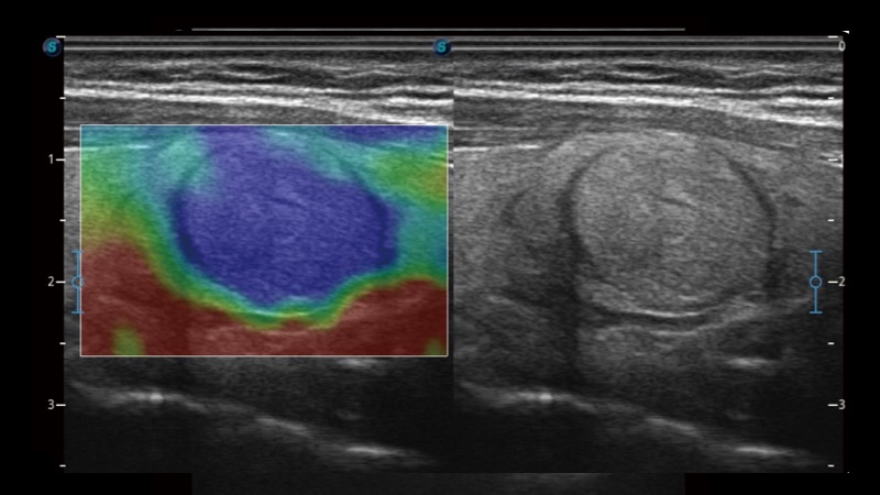
C-xlasto Imaging
C-xlaso imaging enables comprehensive quantitative elastic analysis. Meanwhile, it is supported by multiple probes to ensure good reproducibility and highly consistent quantitative elastic results.
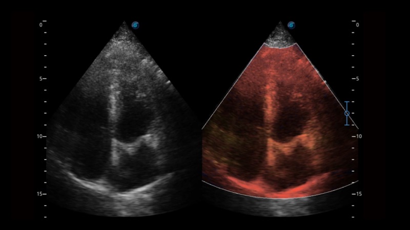
Tissue Doppler Imaging
P12 Elite is endowed with Tissue Doppler Imaging which provides velocities and other clinical information on myocardial functions, facilitating clinical doctors with the ability to analyze and compare the motions of different parts of the patient's heart.
C-xlasto Imaging
C-xlaso imaging enables comprehensive quantitative elastic analysis. Meanwhile, it is supported by multiple probes to ensure good reproducibility and highly consistent quantitative elastic results.
Tissue Doppler Imaging
P12 Elite is endowed with Tissue Doppler Imaging which provides velocities and other clinical information on myocardial functions, facilitating clinical doctors with the ability to analyze and compare the motions of different parts of the patient's heart.
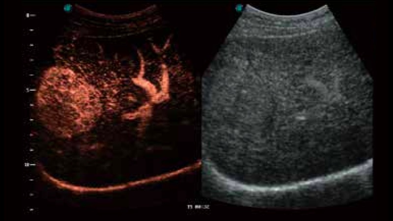
Contrast-Enhanced Ultrasound
The comprehensive contrast-enhance ultrasound imaging and quantification package allow doctors to assess perfusion dynamics in a wide range of clinical setting. The unique Dynamic Acoustic Control technology effectively controls the acoustic pressure of the contrast agent, guaranteeing longer contrast agent duration and better lesion perfusion of delayed phase observation.
C-xlasto Imaging
C-xlaso imaging enables comprehensive quantitative elastic analysis. Meanwhile, it is supported by multiple probes to ensure good reproducibility and highly consistent quantitative elastic results.
Tissue Doppler Imaging
P12 Elite is endowed with Tissue Doppler Imaging which provides velocities and other clinical information on myocardial functions, facilitating clinical doctors with the ability to analyze and compare the motions of different parts of the patient's heart.
Contrast-Enhanced Ultrasound
The comprehensive contrast-enhance ultrasound imaging and quantification package allow doctors to assess perfusion dynamics in a wide range of clinical setting. The unique Dynamic Acoustic Control technology effectively controls the acoustic pressure of the contrast agent, guaranteeing longer contrast agent duration and better lesion perfusion of delayed phase observation.
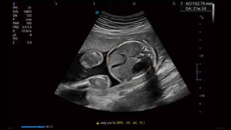
S-Fetus
Based on a big data dependable deep learning algorithm, S-Fetus is a brilliant one-step solution for automatic standard plane acquisition and measurement. With just one click, common fetal biometry results are obtained with high intelligence, accuracy and efficiency, aiming for an unprecedented ease during operation.
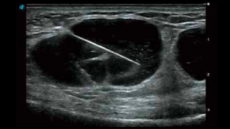
Vis-Needle
Vis-Needle is realized by ultrasound beam steering and deflection. It improves visualization of the needle location in the tissue to minimize harm to the surrounding tissue, increasing the initial success rate and lowering the risk for needle puncture.