


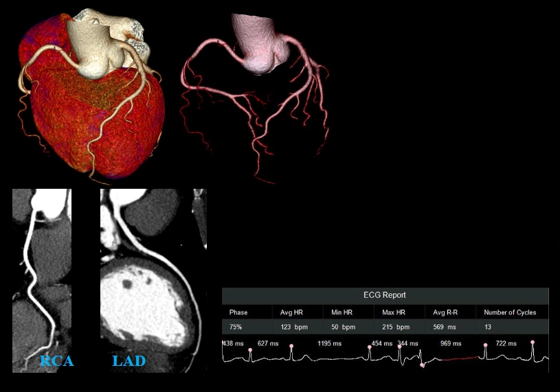


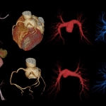
Unprecedented Whole-heart Temporal Resolution

- 16cm Z-Detector
Full heart coverage with a single rotation - 0.25s/rotation
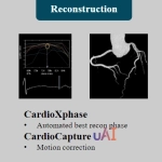
Leading rotation speed at 16cm coverage - CardioCapture uAI
AI empowered motion correction
25ms
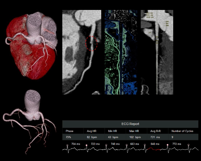
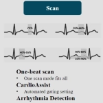
- Along with the full detector coverage and industry leading rotation speed, uCT960+ covers the whole heart in 125ms with half scan.
- The innovative CardioCapturetechnology further boosts the effective whole-heart temporal resolution to 25ms, providing confident diagnostic images for patients with high heart rates and arrhythmias.
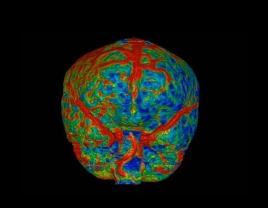
Z-detector Architecture
High Resolution and Ultra-low Noise

16cm Z-coverage
- Full organ coverage in asingle rotation
- 3D ASG manufactured with 3D printing technology for accurate scatter photon shielding
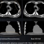
- Real 3D Full reconstruction technology designed to mitigate cone beam artifacts associated with wide coverage systems
Fully Integrated Design
- Innovative Through-silicon-via (TSV)Technology
- cm to μm level signal conducting path shortening
- Uitra-low noise signal output = Low Dose
0.5mm Detector Element Size
- 0.5mm detector acquisition in all FOVs and collimations
- High number of detector elements –299,520
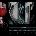
CardioCaptureAI uAI
Empowered Coronary Artery Motion Correction
Why AI?
- Motion correction is essentially the extraction of object movement tracks, based on which the correction is conducted
- The precise estimation of object movement tracks relies on the extraction of coronary arteries and their centerlines
- The conventional vessel extraction is usually based on CT value threshold, fixed coronary models, etc., which often fails especially for vessels with motion artifacts
- The AI based technology learns from different coronary artery images, enabling efficient and precise vessel extraction, showing great advantages for distal vessels
AI Empowered Coronary CTA Workflow
Automated, Standardized and Personalized


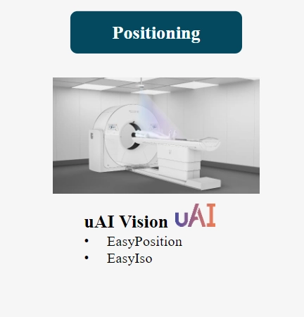
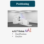
uAIVision
AI Empowered Scan Navigation

- Digitalizing the Patient
Building real-time digital models for every patient with deep learning technology - All Patient Positions
Anatomical structures of the patient can be identified with any positioning - EasyPositioning
Single-click patient positioning with the scan range precisely located based on the protocol selected - EasyIso
Optimize the coronary CTA image quality and patient surface dose distribution
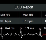
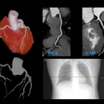
Severe Arrhythmia
50~215 bpmOne-beat Cardiac Scan

- Patient
Female, 58y
Severe arrhythmia with HR 50~215 bpm - CorCTA Axial
Imaging mode 640×0.5mm
0.25s/rotation
Single rotation axial acquisition
100kV
CTDIvol5.0mGy
Effective dose1.13mSv - Contrast
50ml, 370mg/ml, 5ml/s
Atrial Fibrillation 49-165 bpm with Pacemaker
One-beat Cardiac Scan

- Patient
Male, 58y
Atrial Fibrillation with HR 49~165 bpm - CorCTA Axial
Imaging mode 640×0.5mm
0.25s/rotation
Single rotation axial acquisition
100kV
CTDIvol 4.1mGy
Effective dose 0.92mSv - Contrast
50ml, 370mg/ml, 5ml/s
Atrial Fibrillation 43-102 bpm
One-beat Cardiac Scan

- Patient
Male, 71y
Atrial Fibrillation with HR 43~102 bpm - CorCTA Axial
Imaging mode 640×0.5mm
0.25s/rotation
Single rotation axial acquisition
100kV
CTDIvol 4.8mGy
Effective dose1.08mSv - Contrast
55ml, 370mg/ml, 5ml/s
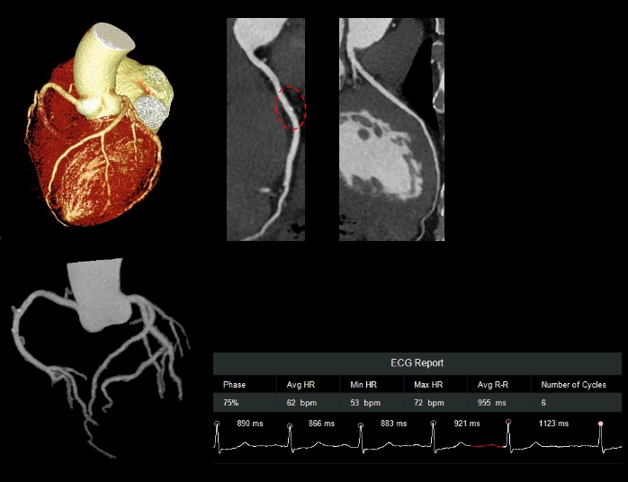
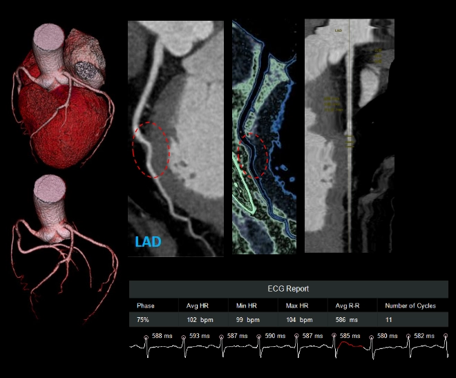
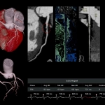
Myocardial Bridging with 102bpm
One-beat Cardiac Scan

- Patient
Female, 73y
102 bpm - CorCTA Axial
Imaging mode 640×0.5mm
0.25s/rotation
Single rotation axial acquisition
100kV
CTDIvol 4.8mGy
Effective dose 1.07mSv - Contrast
45ml, 370mg/ml, 5ml/s
Free-breathing Imaging
One-beat Cardiac Scan

- Patient
Male, 52y
62 bpm
Unable to follow the breathing navigation - CorCTA Axial
Imaging mode 640×0.5mm
0.25s/rotation
Single rotation axial acquisition
100kV
CTDIvol 3.8mGy
Effective dose 0.85mSv - Contrast
40ml, 370mg/ml, 5ml/s
Imaging Challenging Anatomy
One-beat Cardiac Scan


- Patient
Male, 37y
89 bpm
Height 5ft.11in., Weight 287 lbs, BMI 39.2 - CorCTA Axial
Imaging mode 640×0.5mm
0.25s/rotation
Single rotation axial acquisition
120kV
CTDIvol 17mGy
Effective dose 3.88mSv - Contrast
50ml, 370mg/ml, 5ml/s
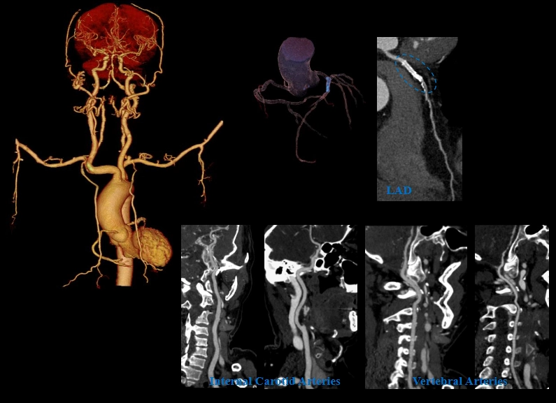
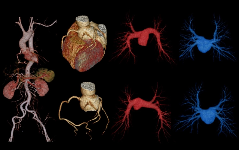
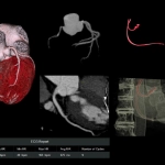
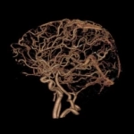
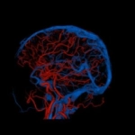
One-stop Cardio-cerebralvascular Imaging
One-beat Cardiac Scan combined with Fast Helical CTA Scan

- Patient
Female, 62y - CorCTA Axial
Imaging mode 640×0.5mm
0.25s/rotation
Single rotation axial acquisition
Effective dose 0.64mSv - Carotid CTA
Imaging mode 320×0.5mm
0.25s/rotation
Scan range 588mm
Effective dose 1.7mSv
Scan Time
4s including the switch from axial to helical scan
Contrast
45ml, 370mg/ml, 5ml/s
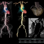
One-stop TAVR Imaging
One-beat Cardiac Scan combined with Fast Helical CTA Scan

- Patient
Female, 74y
49-103bpm - CorCTA Axial
Imaging mode 640×0.5mm
0.25s/rotation
Single rotation axial acquisition
Effective dose 0.91mSv - Aorta CTA
Imaging mode 320×0.5mm
0.25s/rotation - Scan range 650mm
Effective dose 1.75mSv - Scan Time
6s including the switch from axial to helical scan - Contrast
55ml, 370mg/ml, 5ml/s
Enhance the Clinical Decision Support for ER
One-stop Imaging Solutions with Accelerated Workflow
Every second counts in the Emergency Room. CT serves as an important imaging approach for ER due to its capability of fast whole-body scans. uCT960+ is designed to further enhance this capability, providing one-stop imaging solutions with fast and multi-dimensional information for the clinical decision support in ER.
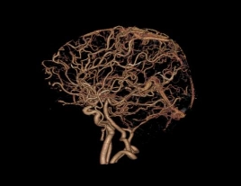
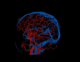
One-stop Stoke Imaging
One Contrast Injection, from Anatomical to Functional Information
Non-contrast CT

- Rule out intracerebral hemorrhage or other non-vascular diseases
- Initial evaluation of cerebral infarction early ischemic clinical signs
CTA

Identify and evaluate the offending vessels for the arterial ischemic stroke (AIS)
4D Dynamic CTA

- Comprehensive analysis of the collateral circulation with multi-phase CTA
- Support analysis for up to 20 time points
Volume CT Perfusion

- Identify the infarction and the salvageable ischemic penumbra
- Adjustable scan interval and dose
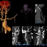
One-stop Triple-rule-out Imaging
One Contrast Injection for the Coronary Artery, Pulmonary Artery/Vein and Aorta CTA

- Patient
Female, 70y
73bpm - Pulmonary CTA Axial
Imaging mode 640×0.5mm
0.25s/rotation
Effective dose 0.9mSv - CorCTA Axial
Imaging mode 640×0.5mm
0.25s/rotation
Single rotation axial acquisition
Effective dose 0.9mSv - Aorta CTA
Imaging mode 320×0.5mm
0.25s/rotation
Scan range 770mm
Effective dose 1.16mSv - Scan Time
6s including the switch from axial to helical scan - Contrast
40ml, 370mg/ml, 5ml/s
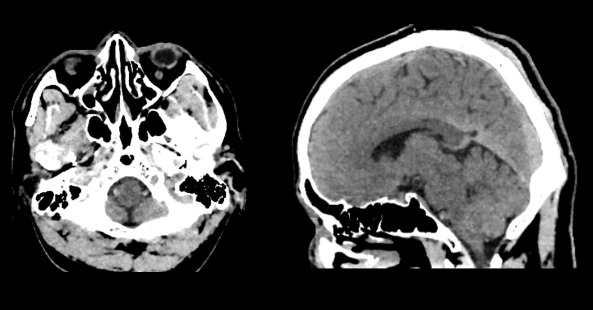
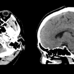
Accelerated Trauma Imaging
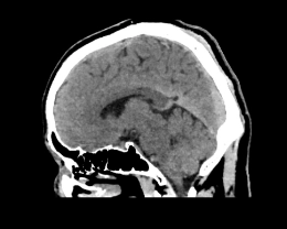
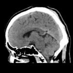
Half-second Full Brain Imaging

Head Axial
Imaging mode 640×0.5mm
0.5s/rotation
Single rotation axial acquisition
120kV, 347mAs
CTDIvol 44.8mGy
- Eliminate motion artifacts caused by involuntary movement
- Clear visualization of the skull base with minimized scatter and cone beam artifacts
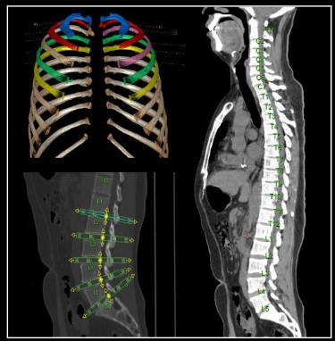
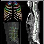
Accelerated Trauma Imaging
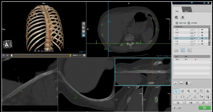
Fast Fracture Localization with the Bone Structure Analysis
Automated rib and spine labeling

Multi-view observation

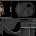
Accelerated Acute Abdomen Imaging
Instant VR/MPR Preview Images

- MPR images can assist the fast localization of lesion, increase the sensitivity and accuracy for the diagnosis of acute abdominal diseases
- Real Time 3D generates VR/MPR preview images along with axial preview images during X-ray exposure
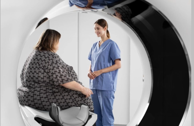
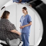
Care for All
Patients and Technologists
Since the beginning of 2020, the COVID-19 pandemic had spread throughout the globe and heavily impacted the healthcare system in a way that never happened before. uCT960+ is designed for safe and precise examinations, both for patients and technologists.
For Elders
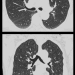
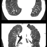
Free-breathing and Motion-free Lung Imaging
74y female with AD Sub-second free-breathing lung imaging

89y male with COPD Sub-second free-breathing lung imaging

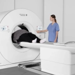
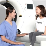
For All Sizes
Improved Patient Experience
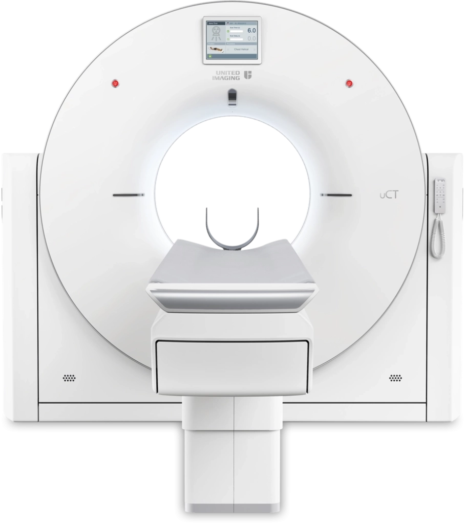
82cm Bore Size

700 lbs Table Load Capacity

For Pediatrics
Dedicated Pediatric Protocols with ALARA Principle

60kV
Scan Mode
Greatly reduces the radiation dose while maintaining the image quality for pediatric patients
16cm
Single-rotation Pediatric Multi-organ Imaging
Reduce the sedation requirement for pediatric patients