TECHNOLOGIES:

Ultra-High-Resolution Digital PET/CT
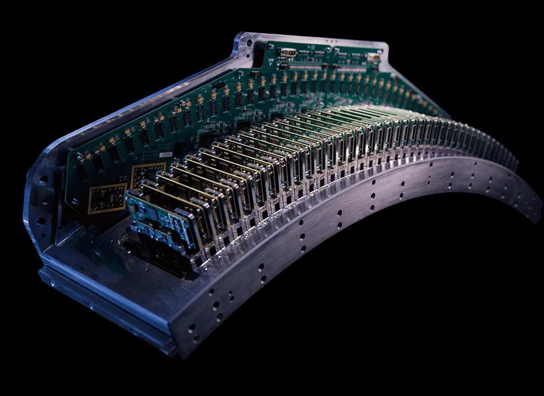
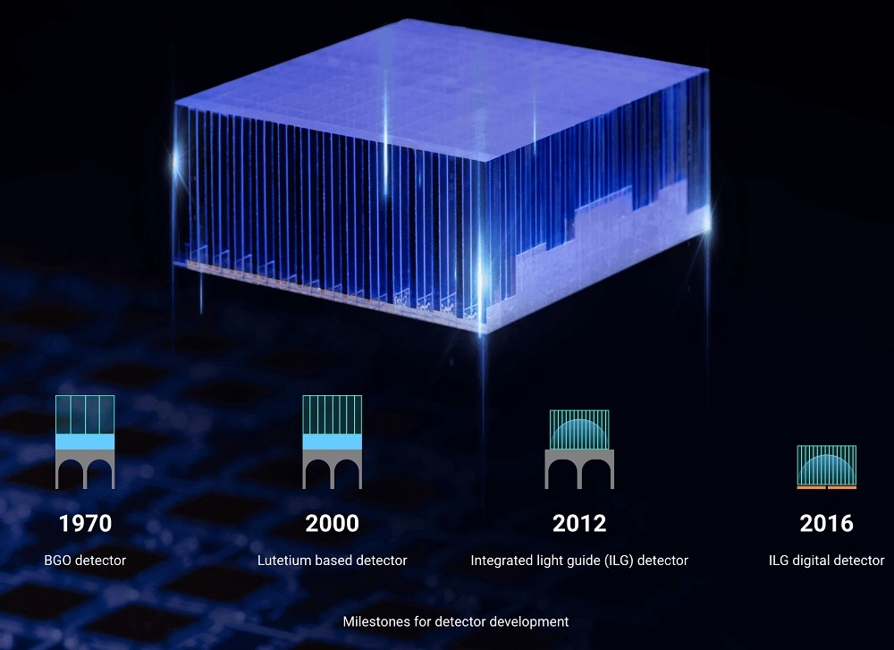
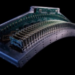
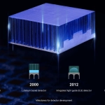
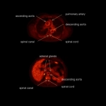
The core of the uEXPLORER PET system is the integrated light guide digital detectors that consist of silicon photomultipliers (SiPMs) and fine 2.76 mm lutetium-yttrium oxyorthosilicate(LYSO) crystals, with over 564,480 crystal elements. This provides exceptional image quality, with 2.9 mm NEMA resolution to improve quantitative accuracy and small lesion detectability.

160-Slice Ultra-Fast CT
The uEXPLORER employs a 160-slice CT that is equipped with the Z-Detector to allow for low dose data acquisition with ultra-low electronic noise. The fast rotation speed enables advanced applications such as cardiac imaging, while the 0.5 mm individual element size allows visualization of very fine structures with every scan. The uEXPLORER provides true clinical versatility without having to choose between fast scans or low dose.

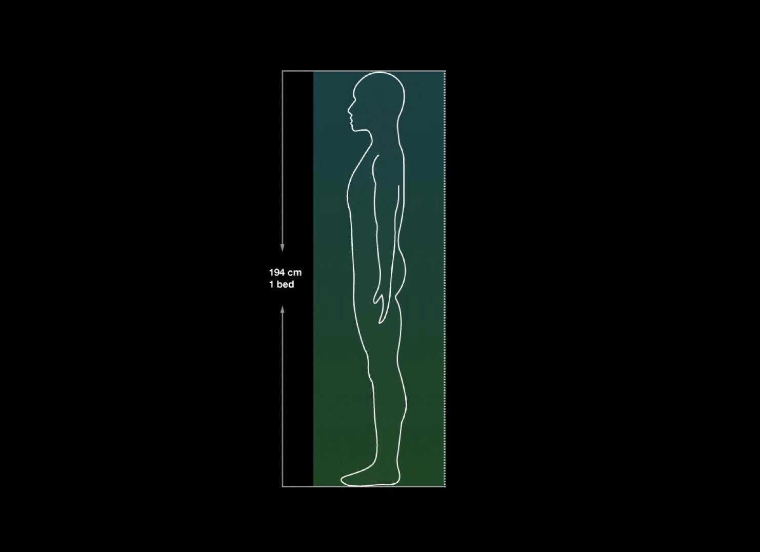
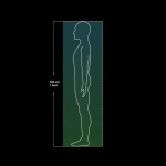
194 cm Axial Field of View (FOV)
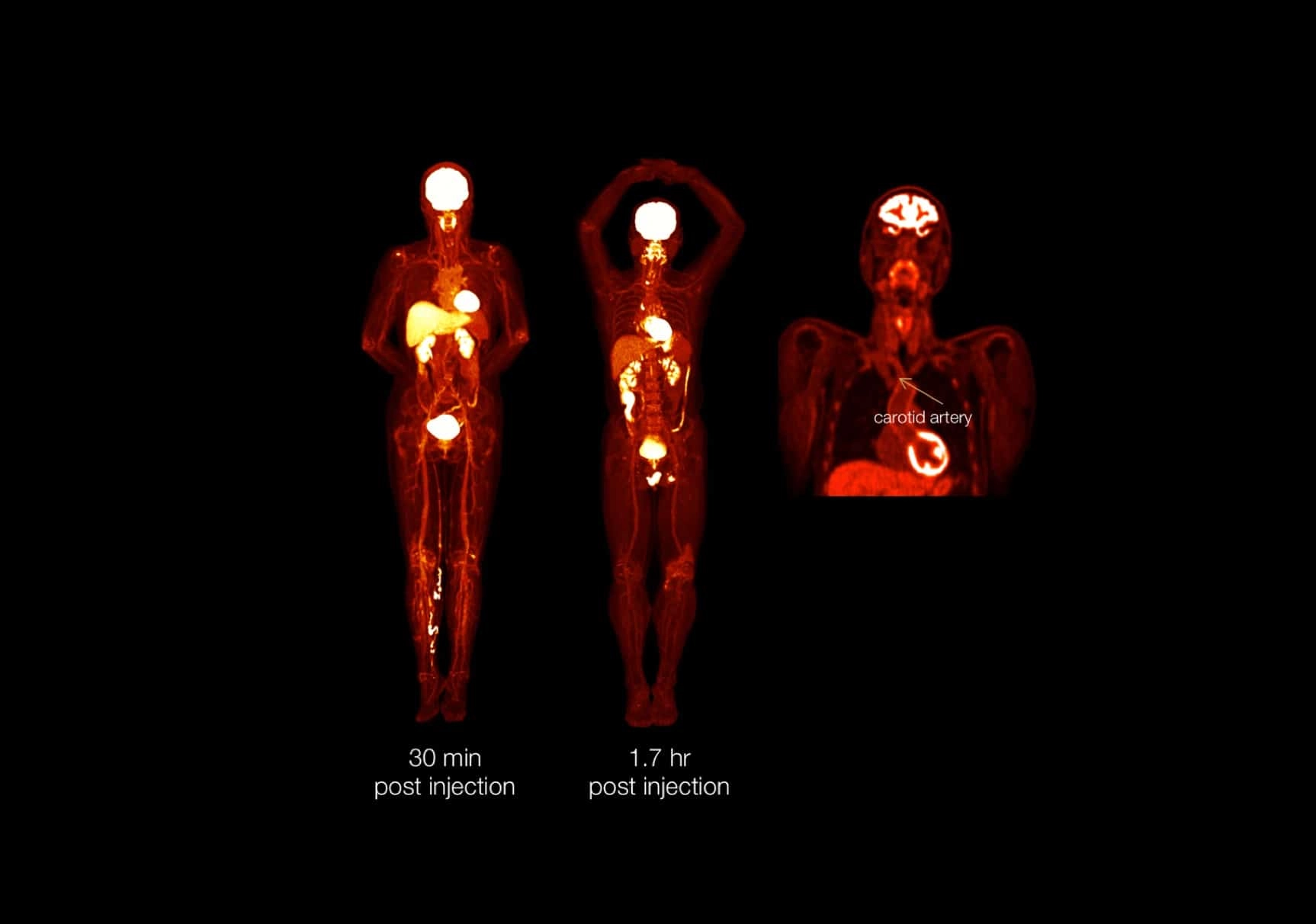
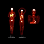
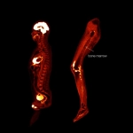
The 194 cm axial PET FOV enables the whole human body to be scanned in one bed position, and powerful computing hardware gives the ability to complete total body reconstruction within three minutes. The uEXPLORER also touts an extremely high sensitivity of 176 cps/kBq, allowing for clinical flexibility with the potential to scan at sub-millicurie tracer dose or in as little time as 30 seconds at standard dose. Together, these core components enable maximum system efficiency, a more effective workflow and an increase in patient throughput.

Clarity for Imaging - Full Field of View Resolutions
Clarity
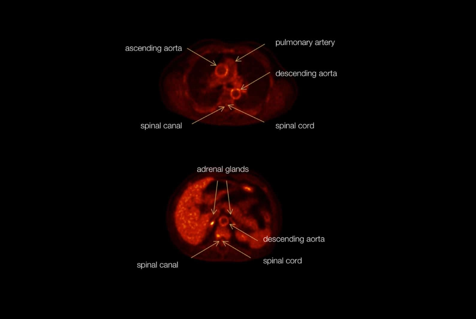
2.9 mm ultra-high NEMA PET resolution improves quantitative accuracy and small lesion detection
Sensitivity
176 cps/kBq high system sensitivity boosts data acquisition and enables sub-millicurie tracer dose
Speed
194 cm axial FOV enables the whole body to be scanned in one bed position

Generate a total body physiological image at a single point in time, in one bed position.

Increase anatomy conspicuity in 18F‑FDG with high sensitivity and the high-resolution digital detectors.

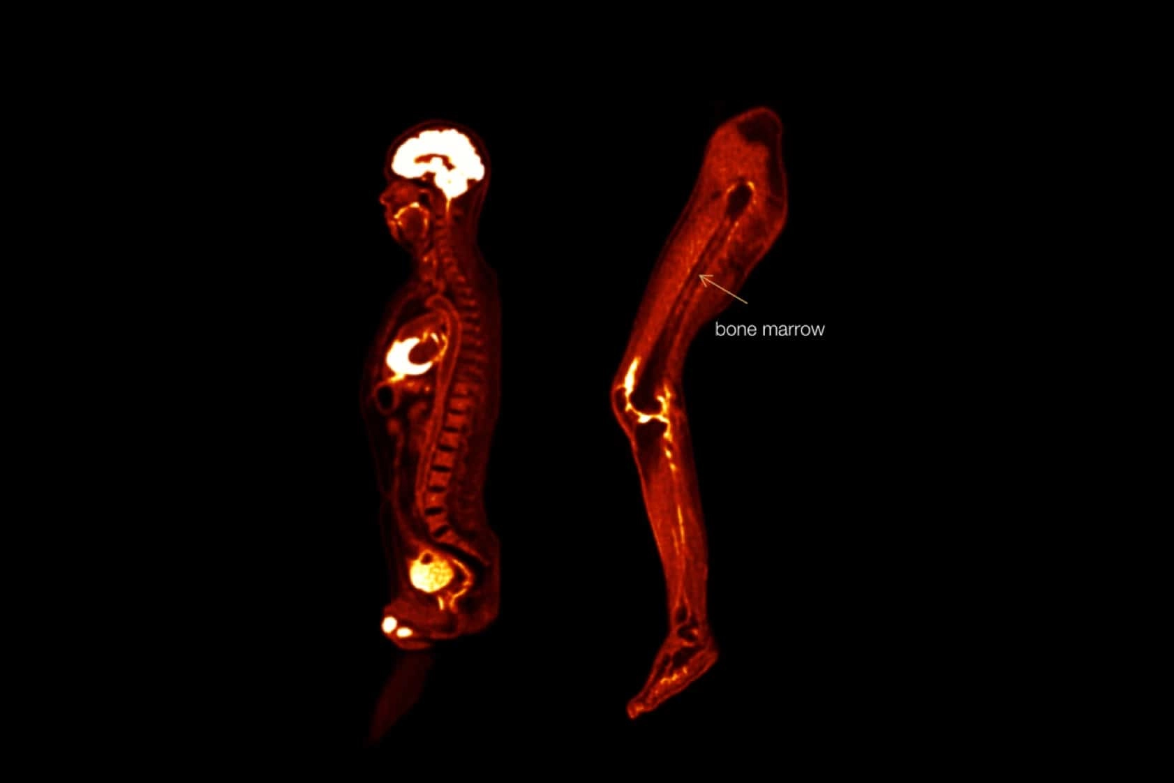
View detailed sagittal and coronal slices with thin axial slices.