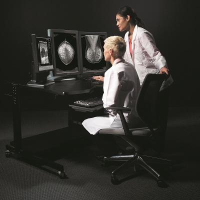
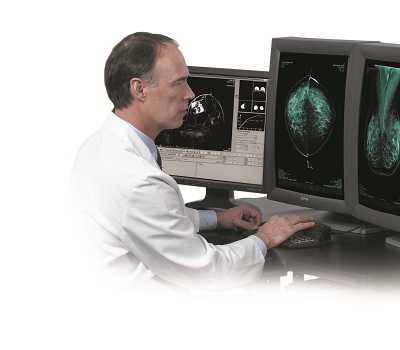
We participate in the Digital Mammography – Tomosynthesis Seminar, providing the diagnostic workstation of the digital mammography unit SELENIA DIMENSIONS.
You can meet us on 2/6/18, at the Choremeio Amphitheater at the Children’s Hospital in Athens, where there will be presentations of incidents of digital mammography, tomosynthesis and contrast enhanced mammography imaging, by experienced doctors/trainers who will participate.
A few words about Hologic Imaging Techniques:


Hologic, a pioneer of the technique of tomosynthesis, has promoted this technique since 2008 and today has more than 8000 world-wide reconfiguration facilities while is the first which has been approved by FDA (DBT / 2D & 3D Image Set)
In particular:
1) It is the first brand which has FDA approval for the superiority of this technique contrary to the classic 2D digital mammography (FDA indication as superior to FFDM).
It is the only one with proven clinical superiority as well as superiority to 2D digital mammography combined with a corresponding reduction in recall rates.
It is the only tomosynthesis technology certified for its superiority contrary to 2D digital mammography, especially for women with dense breasts. “FDA approved as superior to standard 2D mammography for routine breast cancer screening of women with dense breast.”
2) HOLOGIC is the only brand that, throughout independent studies (more than 100), has proven that using SELENIA DIMENSIONS and its cutting-edge technology ensures an increase of the diagnostic sensitivity and specificity of the mammography system, increasing the detection of true cancer findings and simultaneously reducing false cancer findings.
3) HOLOGIC is the only company that with the application of specific tomosynthesis’ technical techniques designed by it for many years, has proven its superiority in 2D & 3D mammography compared to the classic 2D digital mammography.
Particularly, the most recent published studies (indicatively attached) about the use of SELENIA DIMENSIONS digital mammographer for the combination of 2D and 3D digital mammography have proven that we can ensure:
a. 40% increase in the detection of invasive cancers.
b. 27% increase in the detection of cancers (both invasive and non-invasive).
c. 15% reduction in false results.
The evolution of the HOLOGIC tomosynthesis technique is the only FDA approved C-VIEW™ (Synthesized 2D Mammography) Technique.
The revolutionary C-VIEW technology completely eliminates the need for 2D digital mammography, as the 3D synthesis creates the synthesized 2D image of the same breast which as a fully processed image illustrates all the useful information and structure more clearly. Therefore, the overall dose and the time of the full mammogram test, are significantly reduced.
Furthermore, we point out that this specific C-VIEW technique, is the only one FDA approved with clinical studies and clinical appropriate and adequate in order to replace the 2D FFDM digital mammography without requiring 2D digital mammography.
4) SELENIA DIMENSIONS is the only one FDA approved system worldwide that has the pioneering ability to carry out biopsy in 3D imaging that will lead to biopsies and alterations only visible in syntextical images. In addition, due to the fact that through 3D biopsy less exposure is required, we can ensure that the lesions are located easier and the radiation dose to the patient is reduced.
5) It has the pioneering COMBO MODE capability through which 2D and 3D mammography can be captured with a single compression of the breast in order to allow precise comparison and alignment of the findings between shuffle images and normal 2D image.
The combined capture provides the ability to capture 2D images and perform 3D scans at the same compression. This is ideal not only for the technologist, but also for the patients as it reduces the number of compressions, as well as the total time of examination and compression. 2D and 3D scans are taken at the same compression and the image pairs are recorder together. Therefore, any structure at a given coordinate position X, C in the 3D image will depict the same structure in the same X, C region in the 2D image. The study of image pairs, allows the physicians to observe data sets simultaneously.
Furthermore, tomosynthesis, is used in all shots, both in CC and in MLO.
6) CONTRAST ENHANCED MAMOGRAPHY IMAGING (MRI), with which breast images are obtained by contrast injection and the areas of angiogenesis are indicated by increased absorption of contrast agent.


